What Shapes Your Cornea
What Shapes Your Cornea
First of two articles on astigmatism. This one is about the evidence that the cornea changes shape at all. The second, Astigmatism and Focus, is about what you can do with that.
Your cornea changes shape when you read. It changes shape when something presses on your eyelid. And it can be reshaped deliberately, overnight, by a rigid orthokeratology lens worn during sleep — then it changes back once you stop wearing it.
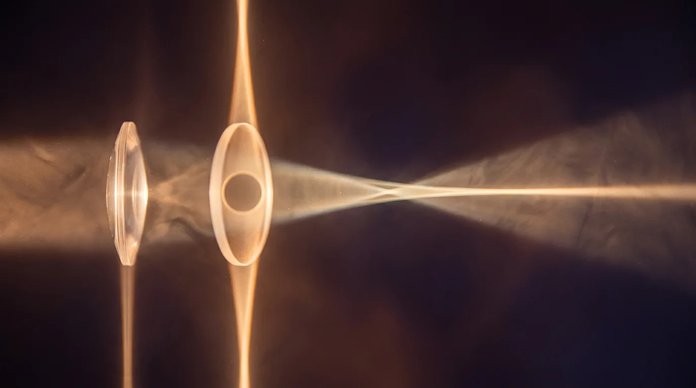
Light through an uneven lens does not meet at a point. It is stretched one way, then the other.
None of that is a fringe claim. All of it has been measured, published, and in one case turned into a product. Yet nobody knows why the curvature of one person's cornea differs from another's — that is not a gap being hidden, it is what the large medical sites say themselves when they reach the question of cause. [1] What is known is that astigmatism does not hold still over a lifetime: it shifts in both amount and direction as people age. [2]
This article is about what has actually been measured.
You don't need to know the mechanism
People got strong lifting weights long before anyone understood protein synthesis. You do not need the mechanism in order to use the effect. If something you do improves what you see, then it improved what you see — whether that happened in the eyelid, in the oblique muscles, or in the cortex.
This matters here, because the mechanism of astigmatism is still being argued about.
Bates blamed the muscles that move the eye. In The Cure of Imperfect Sight by Treatment Without Glasses (1920) he held that refractive errors, astigmatism among them, come from strain in the muscles outside the eyeball — the oblique muscles tightening around it like a tourniquet, the recti pulling it flat against the socket. He went further, and claimed that those same muscles, rather than the lens, are what focus the eye. [3]
That second claim is wrong. Helmholtz, Tscherning and Gullstrand each showed that the lens itself changes shape as the eye focuses, and modern ultrasound imaging has since watched it happen directly. [4] It is worth keeping that failure separate from the first claim, which is a different question and still open.
The accepted account points instead to the shape of the cornea, and to the pressure of the eyelid resting on it. But "accepted" is doing less work in that sentence than you might expect. The most careful review of the question — Read, Collins and Carney, who worked through the theories built on genetics, on extraocular muscle tension, on visual feedback and on eyelid pressure — concluded that no single model of how astigmatism develops has been proven conclusively. [2]
So the argument is open, and the people who know it best say so in print. Nothing that follows depends on closing it. What follows depends on one thing only: that what you see changes, and that you can notice the change.
What has actually been measured
The pressure of the eyelid on the eye is not a guess. It has been measured directly. Researchers at Queensland University of Technology mounted a 0.17 mm tactile piezoresistive sensor on a rigid contact lens and put a number on it: about 8 mmHg of static pressure from the upper lid, resting on the eye. [5] [6]
The more useful evidence, though, is what that pressure does to the shape of the cornea.
- Reading bends the cornea. After fifteen minutes of reading in downward gaze, corneal astigmatism shifts by around a quarter of a dioptre, and it can take up to two hours to return. [7] [8] The effect is larger at a 40° gaze angle than at 20°, because the lid closes further over the cornea. It also depends on what you are doing: reading and microscopy produce larger, more central distortion than computer work, where the gaze is closer to level. The researchers who measured this proposed that lid-induced corneal change may play a part in how myopia develops. [9]
- A lump on the lid creates astigmatism, and removing it takes the astigmatism away. Chalazia larger than 5 mm, sitting centrally on the upper lid, induce measurable astigmatism. Excision changes corneal astigmatism by roughly a third of a dioptre. [10] [11] This is the most convincing evidence available, because the same eye shows you both the onset and the disappearance.
None of these are large numbers. A quarter of a dioptre will not change your life. But they are not zero either, and zero is what you would expect if the shape of your cornea were simply a fact about you.
Try it on yourself
If you have astigmatism, you can take the eyelid out of the equation for a few seconds and watch what happens.
Cover one eye and look at something with fine detail — small print, a distant sign, a line of text across the room. With your free hand, rest a fingertip on the skin of the upper lid, just above the lashes, and lift gently upwards and outwards, so the lid stops resting on the eye. Lift it away. Do not press inwards: the point is to remove pressure, not to add any.
Then let go, and look again.
For some people nothing happens. For others the doubling softens, or sharpens, or leans in a different direction. If it changes, what you have just seen is the number from the start of this section — about eight millimetres of mercury, resting on your eye all day — becoming visible for a moment.
This is the same effect that surgeons produce deliberately. When a drooping eyelid is lifted in surgery, the measured astigmatism of that eye changes with it. [12]
A caution worth taking seriously: clean hands, light touch, and never any pressure on the eye itself. If you have keratoconus, a thin or weakened cornea, or you have had eye surgery recently, skip this — rubbing and pressing are known to make those conditions worse.
The evidence for the eyelid is better than the evidence for the muscles that move the eye. But better is not the same as only. Nobody has shown that the oblique muscles do not contribute. That idea is untested rather than refuted — partly because measuring the force of a muscle wrapped around a globe full of fluid is genuinely hard.
So the honest position is this. Eyelid pressure is documented. A muscular contribution is plausible and unproven. Neither of those sentences needs to be settled before you can start paying attention to what you see.
A lens that reshapes the cornea overnight
There is a treatment in routine clinical use that settles part of this argument, and it has nothing to do with Bates.
Orthokeratology is a rigid lens, made of a highly gas-permeable material, worn while you sleep. It is built with a reverse geometry, so that overnight it flattens the centre of the cornea. In the morning you take it out and you see clearly, without glasses, for the rest of the day. Most of the change happens after the very first night, and the effect settles within seven to ten days. Stop wearing it and the cornea returns to what it was. [13]
It works on astigmatism too. Toric designs, built for eyes that have it, reduce astigmatism by around four fifths. [14]
Stay with what that means for a moment. A piece of plastic, pressing gently for a few hours a night, changes the shape of the cornea enough to make glasses unnecessary — and the change undoes itself once the pressure is taken away.
Where that leaves you
Put the measurements side by side and one thing is consistent. The cornea answers to what is done to it, and what it answers with is reversible. Reading bends it for an hour or two. A lump on the lid bends it for as long as the lump is there. A rigid lens bends it overnight, and goes on bending it for as long as you keep wearing it.
So the shape of your cornea is not quite a fact about you. It is closer to a running total of what has been pressing on your eye.
Which raises the question this article cannot answer. If the shape is not fixed, is there anything you can do about it yourself — and how would you ever know whether it had worked?
The next article takes that up: Astigmatism and Focus.
References
[1] Astigmatism: symptoms and causes. Mayo Clinic.
[2] Read, S. A., Collins, M. J., & Carney, L. G. (2007). A review of astigmatism and its possible genesis. Clinical and Experimental Optometry, 90(1), 5-19.
[3] Bates, W. H. (1920). The Cure of Imperfect Sight by Treatment Without Glasses. Central Fixation Publishing Company.
[4] Mechanism of accommodation: a review of theoretical propositions. African Vision and Eye Health.
[5] Shaw, A. J. (2009). Eyelid pressure on the cornea. PhD thesis, Queensland University of Technology.
[6] Shaw, A. J., Collins, M. J., Davis, B. A., & Carney, L. G. (2009). A technique to measure eyelid pressure using piezoresistive sensors. IEEE Transactions on Biomedical Engineering.
[7] Collins, M. J., et al. (2005). Regression of lid-induced corneal topography changes after reading. Optometry and Vision Science.
[8] Niyazmand, H., et al. (2013). Anterior segment changes following short-term reading and its correlation with corneal biomechanical characteristics. Ophthalmic and Physiological Optics.
[9] Collins, M. J., et al. (2006). Corneal optics after reading, microscopy and computer work. Acta Ophthalmologica Scandinavica.
[10] Bagheri, A., Hasani, H. R., Karimian, F., Abrishami, M., & Yazdani, S. (2009). Effect of chalazion excision on refractive error and corneal topography. European Journal of Ophthalmology, 19(4).
[11] Jin, K. W., Shin, Y. J., & Hyon, J. Y. (2017). Effects of chalazia on corneal astigmatism. BMC Ophthalmology, 17(1).
[12] Characteristics of astigmatism before and 1 month after blepharoptosis surgery in patients with acquired ptosis. PLOS ONE (2021).
[13] Swarbrick, H. A. (2006). Orthokeratology review and update. Clinical and Experimental Optometry, 89(3).
[14] Clinical safety and efficacy of orthokeratology contact lenses with toric peripheral curves: a review of the literature.